By Dr.Shahzad khurram , Verified medical authority
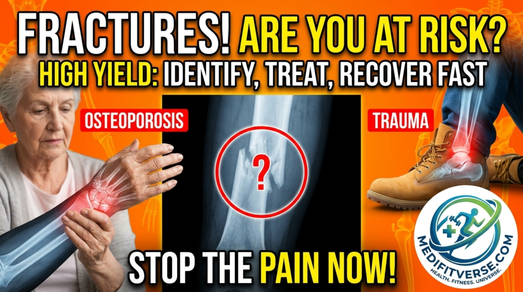
If you’ve ever stared at an X-ray during rounds and blanked on whether it’s a Colles’ or a Smith’s fracture, you’re not alone. Fracture types confuse medical students, physiotherapy trainees, and even busy residents — not because the concepts are hard, but because nine similar sounding names get thrown at you all at once.
This guide breaks down the 9 fracture types that show up again and again on the USMLE, COMLEX, and in real clinical practice. Think of it as the reference sheet you wish someone had handed you on day one of orthopedics rotation.
#Orthopedics
Why These 9 Fractures Matter
These aren’t random topics. Each one teaches a core orthopedic principle, mechanism of injury, nerve or vascular risk, and the management decision point between casting and surgery. Examiners love them because a single clinical vignette (a fall, a punch, a car accident) can test whether you understand anatomy, mechanism, and treatment all at once.
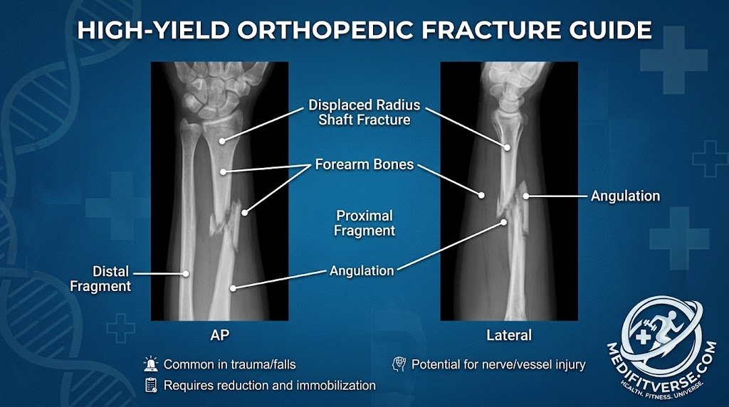
Let’s go through them one at a time. Feel yourself as an orthopedics.
1. Colles’ Fracture
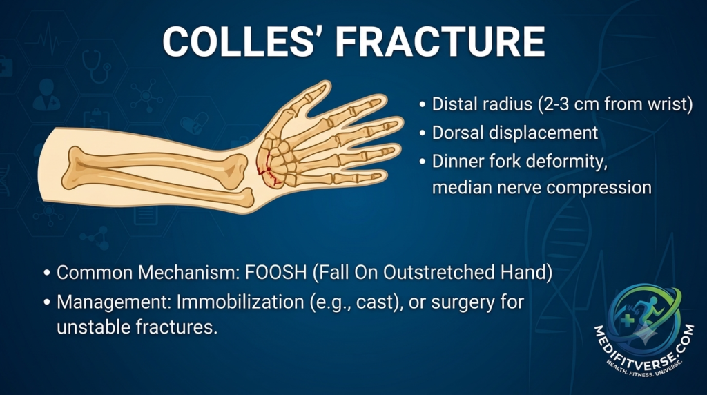
- Location: Distal radius, roughly 2–3 cm from the wrist joint
- Displacement: Dorsal (backward)
- Classic sign: “Dinner fork” deformity; watch for median nerve compression
- Mechanism: FOOSH — Fall On Outstretched Hand
- Management: Closed reduction and cast immobilization for stable fractures; surgical fixation for unstable or comminuted patterns
Clinical Pearl
This is the most common wrist fracture in adults, especially postmenopausal women with osteoporosis. If a patient reports tingling in the thumb, index, and middle fingers, orthopedics always think acute carpal tunnel from median nerve involvement.
2. Smith’s Fracture
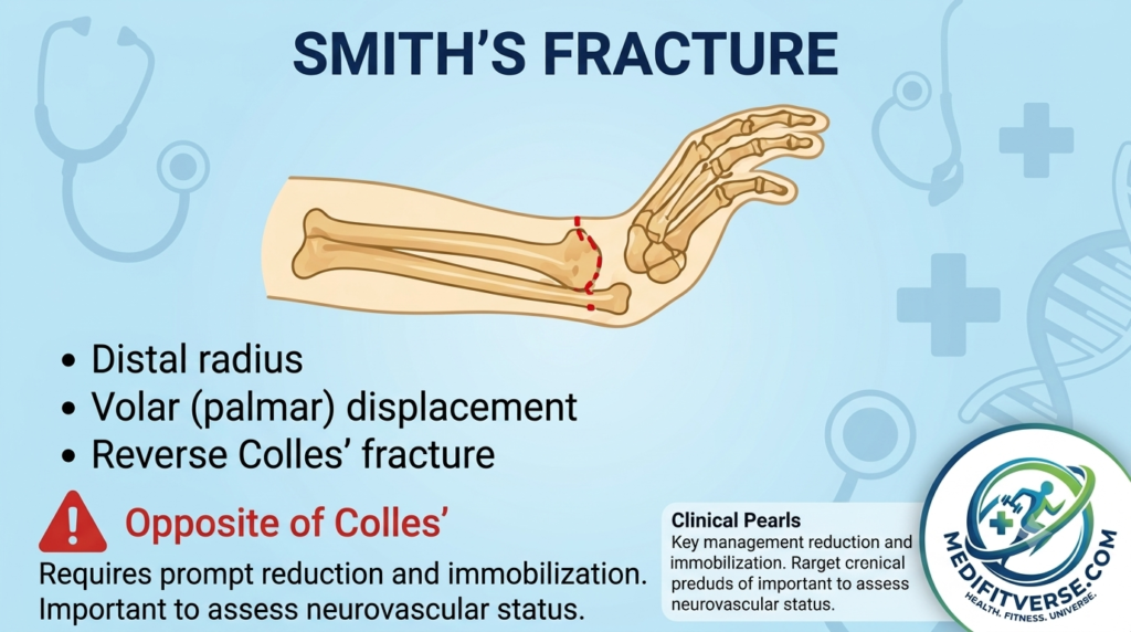
- Location: Distal radius
- Displacement: Volar (palmar) the opposite of Colles’
- Also called: Reverse Colles’ fracture
- Mechanism: Fall onto a flexed wrist, or a direct blow to the back of the wrist
- Management: Requires prompt reduction and immobilization; neurovascular status must be checked carefully
Clinical Pearl
Students often mix up Colles’ and Smith’s. A simple memory trick: Colles’ = Cocked back (dorsal), Smith’s = the opposite “Orthopedics mnemonic”.
3. Monteggia Fracture
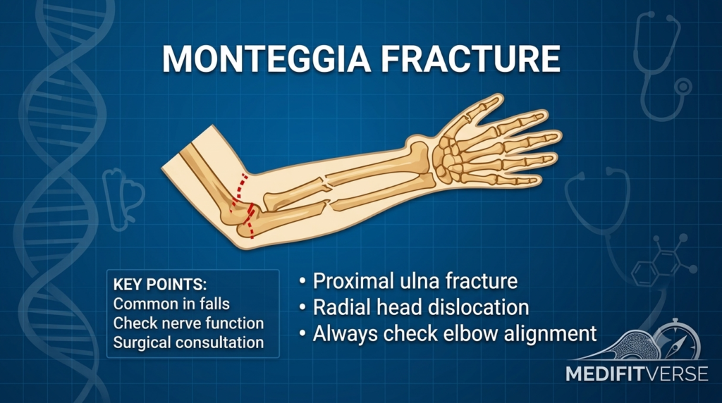
- Injury pattern: Proximal ulna fracture plus radial head dislocation
- Common cause: Falls, especially in children
- Key exam step: Always check elbow alignment and distal nerve function
- Management: Surgical consultation is typically needed to restore both bone alignment and joint stability
Clinical Pearl
“Monteggia” and “ulna” both start with vowel-adjacent sounds if you say them fast, a silly trick, but it helps some students remember that the ulna is the fractured bone here, with the radius dislocating. Orthopedics opinion needed here.
4. Galeazzi Fracture
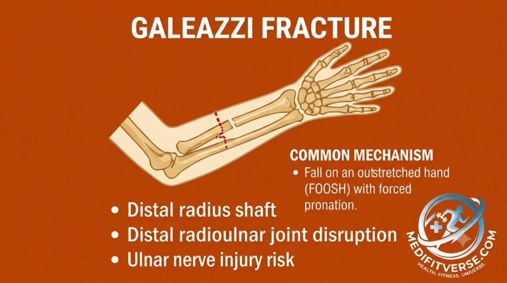
- Location: Distal radial shaft fracture
- Associated injury: Distal radioulnar joint (DRUJ) disruption
- Risk: Ulnar nerve injury
- Mechanism: FOOSH with forced pronation
Clinical Pearl
A helpful orthopedics contrast: Monteggia affects the ulna (proximal) with radial head dislocation; Galeazzi affects the radius (distal) with DRUJ disruption. Students often remember this as “MUGR” — Monteggia = Ulna, Galeazzi = Radius.
5. Boxer’s Fracture
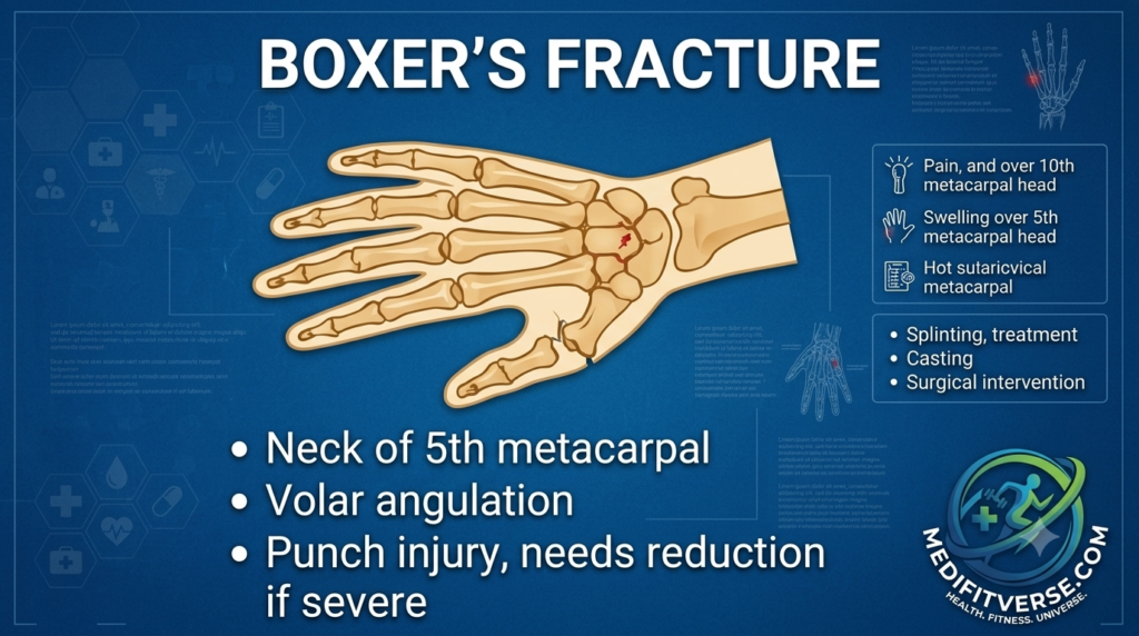
- Location: Neck of the 5th metacarpal
- Displacement: Volar angulation
- Mechanism: Punch injury (closed fist striking a hard surface)
- Symptoms: Pain and swelling over the 5th metacarpal head, tenderness on palpation
- Management: Splinting for mild angulation; reduction or surgical fixation if the deformity is severe
Clinical Pearl
Always ask about the mechanism directly, patients are sometimes reluctant to admit they punched a wall or another person. Also check for “fight bite” wounds over the knuckles, which carry a high infection risk if skin was broken on contact with teeth.
6. Bennett’s Fracture
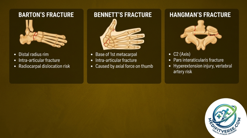
- Location: Base of the 1st metacarpal
- Type: Intraarticular fracture
- Mechanism: Axial force applied to a flexed thumb (e.g., punching with the thumb tucked incorrectly)
- Management: Because it’s intraarticular and unstable, it often needs surgical fixation to prevent longterm joint problems
Clinical Pearl
Don’t confuse this with Boxer’s fracture. Bennett’s involves the thumb’s base and the joint surface, which makes it inherently unstable and much more likely to need surgery than a simple Boxer’s fracture.
7. Barton’s Fracture
- Location: Distal radius rim
- Type: Intraarticular fracture
- Risk: Radiocarpal dislocation
- Management: Often unstable; frequently requires open reduction and internal fixation (ORIF)
Clinical Pearl
Barton’s is essentially a Colles’ or Smith’s fracture that extends into the joint surface, which is exactly why it behaves differently, the joint involvement makes it prone to instability even after reduction.
9. Hangman’s Fracture
- Location: C2 (the axis)
- Pattern: Pars interarticularis fracture
- Mechanism: Hyperextension injury, such as in high speed motor vehicle collisions
- Risk: Vertebral artery injury
- Management: Immobilization is standard for stable patterns; surgery is reserved for significant displacement or instability
Clinical Pearl
The name comes from the injury pattern seen historically in judicial hangings, but today it’s most often seen in high-speed car accidents. Don’t let the dramatic name distract you from checking vertebral artery flow on imaging.
8. Jefferson Fracture
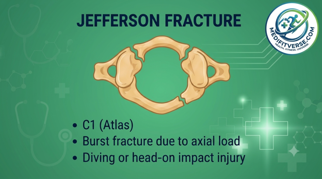
- Location: C1 (the atlas)
- Pattern: Burst fracture caused by axial load
- Common mechanism: Diving into shallow water or a head on impact
- Management: Immobilization with a rigid collar or halo device; neurosurgical or spine surgery consultation is essential given the proximity to the spinal cord
Clinical Pearl
Always assume cervical spine instability until imaging clears it. Any patient with a high energy axial load mechanism (diving, a fall onto the head) needs full spinal precautions before you even get to the X-ray.
Quick-Reference Comparison Table
| Fracture | Bone/Level | Key Mechanism | Distinguishing Sign | Typical Management |
|---|---|---|---|---|
| Colles’ | Distal radius | FOOSH | Dorsal (“dinner fork”) deformity | Cast; ORIF if unstable |
| Smith’s | Distal radius | Fall on flexed wrist | Volar displacement | Reduction + immobilization |
| Monteggia | Proximal ulna | Fall/direct trauma | Radial head dislocation | Surgical consult |
| Galeazzi | Distal radial shaft | FOOSH with pronation | DRUJ disruption | Surgical consult |
| Boxer’s | 5th metacarpal neck | Punch injury | Volar angulation | Splint; reduction if severe |
| Bennett’s | Base of 1st metacarpal | Axial force on thumb | Intra-articular fracture | Usually surgical |
| Barton’s | Distal radius rim | FOOSH | Radiocarpal dislocation risk | Often ORIF |
| Jefferson | C1 (atlas) | Axial load (diving) | Burst pattern | Collar/halo; spine consult |
| Hangman’s | C2 (axis) | Hyperextension | Pars interarticularis fracture | Immobilization or surgery |
How to Actually Remember These for the Boards
- Group by region first, mechanism second. Wrist fractures (Colles’, Smith’s, Barton’s), forearm fractures with joint involvement (Monteggia, Galeazzi), hand fractures (Boxer’s, Bennett’s), and cervical spine fractures (Jefferson, Hangman’s).
- Draw it, don’t just read it. Sketching the direction of displacement (dorsal vs. volar) cements the difference between Colles’ and Smith’s far better than memorizing definitions.
- Attach a mechanism story. A punch, a dive, a fall on an outstretched hand, this style questions almost always test mechanism recognition first.
- Always ask “does this need surgery?” Nearly every board question ends with a management decision, so know which fractures are inherently unstable (Bennett’s, Barton’s, Monteggia, Galeazzi) versus typically manageable with casting (simple Colles’, stable Boxer’s).
A Note on Sources
This overview is meant as a study aid, not a replacement for your institution’s clinical guidelines. For deeper reading and case based learning, reputable references include:
- AAP (American Academy of Pediatrics) for pediatric-specific fracture patterns, especially relevant to Monteggia fractures in children
- https://pubmed.ncbi.nlm.nih.gov/9682084/
- PubMed / NCBI for peer reviewed orthopedic literature
- NHS.uk for patient-facing fracture management overviews
- Medscape for clinical reference summaries on individual fracture types
- CDC.gov for fall-related injury and osteoporosis fracture risk data
Always cross check specifics against your local clinical protocols and current edition textbooks, since management (especially surgical thresholds) can vary by institution.
Final Thoughts
According to me nine fractures, nine stories, one shared lesson: orthopedics rewards pattern recognition. Once you’ve connected each type to its mechanism and its management decision, these stop being memorization tasks and start being clinical reasoning, which is exactly what boards, and real patients, are testing for.
What is the difference between a Colles’ and a Smith’s fracture?
A Colles’ fracture involves dorsal displacement of the distal radius.
A Smith’s fracture is the “reverse” of a Colles’, characterized by volar (palmar) displacement.
What defines a Monteggia fracture?
It is an injury involving a proximal ulna fracture. It is accompanied by a radial head dislocation.
Why is a Jefferson fracture considered serious?
It is a burst fracture of the C1 (Atlas) vertebra. It is typically caused by a high-impact axial load, such as diving or a head-on impact
Dr. Shahzad Khurram is a dedicated Pediatrician and medical researcher passionate about empowering parents with evidence-based health insights. With a clinical focus on childhood nutrition and neurodevelopment, Dr. Khurram translates latest clinical data into actionable advice for modern families. Through Medifitverse, he bridges the gap between pediatric science and everyday parenting, ensuring every child has the nutritional foundation they need to thrive. His work is guided by the latest standards from the American Academy of Pediatrics and current nutritional research.
Medical Disclaimer: The information on Medifitverse, including text, graphics, and images, is for educational and informational purposes only. This content is not intended to be a substitute for professional medical advice, diagnosis, or treatment. Always seek the advice of your pediatrician or other qualified health provider with any questions you may have regarding a medical condition. Never disregard professional medical advice or delay in seeking it because of something you have read on this website.However, every baby is different. Consult your doctor before introducing allergens or changing your infant’s diet.
NOTE: Check my folic acid for kids blog which helps autistic kids.
Contact Dr. Shahzad Khurram here, or click whatsapp icon for direct chat.